Functional MR (fMRI) imaging shows structural and functional alterations in the brains of people with opioid use disorder, according to a team of researchers from Yale School of Medicine in New Haven, CT.
The study results could improve treatment for those who suffer from opioid use disorder, wrote a team led by Saloni Mehta, MBBS. The findings were published December 10 in Radiology.
"We observed widespread increases in global connectivity in individuals with [the disease]," Mehta said in a statement released by the RSNA. "Our goal is to better understand what could have caused these alterations to inform new treatment targets."
Opioids include synthetic drugs such as fentanyl, prescription pain relievers such as oxycodone, and illegal narcotics such as heroin. They are a significant contributor to drug overdoses: In fact, the U.S. Centers for Disease Control and Prevention's (CDC) National Center for Health Statistics estimates that there were 81,083 overdose deaths involving opioids in the U.S. last year.
"We are in the midst of an opioid epidemic, with millions affected worldwide and more than 80,000 deaths related to opioid overdoses in the U.S. last year alone," Mehta said. "We need to get a better understanding of the system-level neural alterations associated with opioid use disorder."
To do this, Mehta and colleagues analyzed data from structural MRI and fMRI exams taken from the National Institutes of Health-funded Collaboration Linking Opioid Use Disorder and Sleep study (CLOUDS); the exams were performed between February 2021 and May 2023. There were 103 structural MRI studies from individuals with opioid use disorder and 105 from the control group, as well as resting state fMRI data from 74 participants with opioid use disorder and from 100 controls.
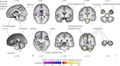
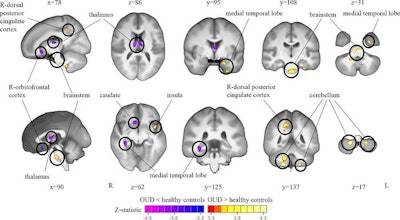
The investigators found that, in people with opioid use disorder, the thalamus and right medial temporal lobe of the brain were smaller in volume, while the cerebellum and brainstem were larger in volume than in controls -- and that these brain regions had higher functional connectivity in individuals with the disease compared with controls. The group also reported that women with opioid use disorder had smaller medial prefrontal cortex volume compared with male counterparts.
 Tensor-based morphometry (TBM) analysis of T1-weighted MRI scans shows a comparison of brain volumes in participants with opioid use disorder (OUD) and healthy control participants. Widespread volume differences are observed between participants with OUD and healthy controls when accounting for total brain volume. Specifically, the bilateral thalamus, right caudate and orbitofrontal cortex, and right medial temporal lobe show lower volume in participants with OUD compared with healthy controls. The left medial temporal lobe, brainstem, bilateral cerebellum, left insula, and right dorsal posterior cingulate cortex show greater volume in those with OUD compared with healthy controls. All results are shown at p
The study "opens avenues for exploring the potential effects of specific interventions that may help to mitigate or even reverse the observed structural and functional changes in the brain," wrote Prof. Dr. Massimo Filippi and Roberta Messina, MD, PhD, both of Vita-Salute San Raffaele University in Milan, Italy, in an accompanying commentary.
"Such interventions could be important in promoting recovery and improving the overall outcomes for individuals with opioid use disorder," they wrote.
The complete study can be found here.

Whether you are a professional looking for a new job or a representative of an organization who needs workforce solutions - we are here to help.