
PET/CT scans with an experimental radiotracer that targets programmed death ligand 1 (PD-L1) protein on cancer cells can reveal the full burden of disease in patients with non-small cell lung cancer, according to a study published August 2026 in Radiology.
A group from the First Affiliated Hospital of Xiamen University in Xiamen, China, performed gallium-68 (Ga-68) NK224 PET/CT scans in participants with newly diagnosed, recurrent, or metastatic non-small cell lung cancer (NSCLC), with findings suggesting the approach can overcome inherent sampling limitations of single-site biopsies. Ultimately, the modality may aid personalized immunotherapy strategies for patients with multifocal metastatic disease, the authors suggested.
“Ga-68 NK224 PET/CT enabled whole-body assessment of programmed death ligand 1 heterogeneity in participants with non–small cell lung cancer and revealed lesion-level heterogeneity not captured through biopsies,” noted lead authors Liang Zhao, MD, and Hui Zhou, MD, and colleagues. “Ga-68 NK224 uptake was associated with immunotherapy response.”
Currently, PD-L1 immunohistochemical analysis performed on biopsy specimens is the standard method for evaluating patients for immunotherapy. However, increasing evidence indicates that PD-L1 expression exhibits pronounced intratumoral and intertumoral heterogeneity, which can make immunotherapy decision-making based on biopsies challenging, the researchers explained.
Alternatively, molecular imaging can provide whole-body visualization and quantitative assessment of PD-L1 expression, and to that end the group previously developed Ga-68 NK224 and found it safe and effective in a small group of patients. In this study, the researchers tested it in a larger cohort in a prospective study from December 2023 to July 2025.
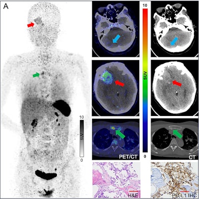
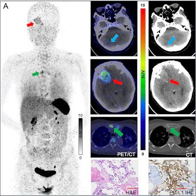 (A) Ga-68 NK224 PET/CT maximum intensity projection image (left), axial PET/CT images (center), and axial CT images (right) in a 43-year-old female participant with lung adenocarcinoma show lesions in the right cerebellum (blue arrow), right frontal lobe (red arrow), and sternum (green arrow) with low, intermediate, and high tracer uptake, respectively. Photomicrographs (scale bar, 100 μm) with hematoxylin-eosin (H&E) staining (center bottom) and programmed death ligand 1 (PD-L1) immunohistochemical (IHC) staining (right bottom) show high PD-L1 expression in the sternal lesion (tumor proportion score ≥ 50%). RNSAAccording to the findings, Ga-68 NK224 uptake differed significantly across PD-L1 tumor proportion score (TPS) categories (based on the percentage of positive tumor cells in the biopsy stain) using both PET biopsy-plane (local) and whole-lesion regions of interest (ROIs) (both p
Intratumoral heterogeneity was evident, with greater Ga-68 NK224 uptake variability in whole-lesion ROIs than biopsy-plane ROIs (median SUVmax 1.0 versus 0.4; p
“By overcoming the inherent sampling limitations of single-site biopsy, Ga-68 NK224 PET/CT provides a more comprehensive assessment of tumor immune phenotypes,” the authors wrote.
Validating quantitative uptake thresholds and determining whether PD-L1 PET-guided treatment selection improves outcomes will require larger multicenter and multiethnic trials, they concluded.
Read the full study here.
Whether you are a professional looking for a new job or a representative of an organization who needs workforce solutions - we are here to help.