Researchers using quantitative susceptibility mapping (QSM) MRI have discovered evidence of the long-term effects of severe COVID-19 on the brain. Their findings were published October 7 in the journal Brain.
A team led by Catarina Rua, MD, of the University of Cambridge in the U.K., reported brain imaging findings on MRI in COVID-19 survivors who had been hospitalized. These findings included increased susceptibility (that is, magnetization) in the medulla, pons, and midbrain regions of the brainstem, which translated to worse disease severity and functional recovery outcomes as well as higher acute inflammatory markers.
"Damage to the brainstem -- the brain's 'control center' -- is behind long-lasting physical and psychiatric effects of severe COVID-19 infection," the group noted in a statement released by the university.
Postmortem studies have shown that patients with acute COVID-19 manifest pathological changes in their central nervous system, specifically the brainstem; the hypothesis is that these changes are caused by para- or postinfection immune responses, the team explained. But brainstem involvement identified on MRI after COVID-19 recovery has remained inconclusive.
In an effort to clarify the long-term effects of COVID-19 on the brain, Rua and colleagues conducted a study that included 30 patients who underwent a 7-tesla quantitative susceptibility mapping MRI exam between 93 and 548 days after hospital admission for the disease; the patients had been admitted to the hospital with severe COVID-19 before vaccines were available. These individuals' imaging results were compared to 51 age-matched controls without prior history of COVID-19 who also underwent this type of MR imaging.
The investigators correlated study participants' QSM signals with the following markers:
Disease severity (measured by duration of hospital admission and COVID-19 severity scale)Inflammatory response during the acute illness (via C-reactive protein, D-dimer, and platelet level results) Functional recovery (using a modified Rankin scale) Depression (Patient Health Questionnaire-9) Anxiety (Generalized Anxiety Disorder-7)Overall, the group found increased MRI susceptibility in the inferior medullary reticular formation and the raphe pallidus and obscurus in patients who had recovered from severe COVID-19 compared to the healthy controls. These regions are associated with symptoms such as breathlessness, fatigue, and anxiety.
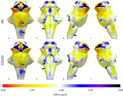
3D projections of the QSM χ maps on the rendered brainstem ROI extracted from the FreeSurfer segmentation for the healthy control group and the COVID group. The COVID group shows increased χ in the brainstem, specifically in the medulla and pons (black arrows). Images courtesy of the University of Cambridge.
"Our key findings were that in COVID-19 survivors, multiple regions of the medulla oblongata, pons, and midbrain show magnetic resonance susceptibility abnormalities at a median time of 6.5 months from hospital admission," the group wrote. "These differences are consistent with a neuroinflammatory response. The fact that these regions that were affected several weeks after hospitalization are the sites of respiratory pathways suggests that lasting symptoms might be an indirect effect of brainstem inflammatory injury following COVID-19."
The researchers hope that the study results will contribute to a better understanding of the long-term effects of severe COVID-19, according to study co-author James Rowe, PhD, also of the university.
"The brainstem is the critical junction box between our conscious selves and what is happening in our bodies," he said in the statement. "The ability to see and understand how the brainstem changes in response to COVID-19 will help explain and treat the long-term effects more effectively."
The complete study can be found here.

Whether you are a professional looking for a new job or a representative of an organization who needs workforce solutions - we are here to help.