
A physics-based technique appears to harmonize CT images to a reference quality index, thus boosting the reproducibility of lung density measurements, researchers have reported.
A team led by Saman Sotoudeh-Paima, PhD, of Duke University School of Medicine in Durham, NC, found that compared with the traditional approach for assessing lung density (MF-VALD, or Median Filtering followed by Volume-Adjusted Lung Density), its "image harmonization pipeline" approach demonstrated better performance. The study results were published March 12 in Radiology: Cardiothoracic Imaging.
"Our image harmonization pipeline reduced variations in lung density measurements," the authors noted.
CT is increasingly used for disease assessment, but using it to quantify lung density remains a challenge -- influenced by differences in lung volumes, CT equipment, radiation dose levels, and image reconstruction protocols, the authors wrote, noting that "image harmonization is critically needed to enable robust and reliable quantification of lung density across varying conditions."
Sotoudeh-Paima and colleagues developed a "sequential physics-based harmonization pipeline" that targeted three principal sources of variability: spatial resolution, image noise, and lung volume. (A sequential physics-based harmonization pipeline is an image processing framework that uses physical properties of CT imaging to correct for sources of measurement variability.) For their study, they used data from 1,159 individuals who participated in the COPDGene study; all underwent same-day full-dose (200 mAs) and reduced-dose (40 mAs to 80 mAs) chest CT imaging between November 2014 and July 2017. The team tracked these sources of variability and also calculated the percentage of lung voxels less than -950 Hounsfield units (HU) and the 15th percentile (Perc15) of a lung density histogram that the algorithm produced.
The group reported the following:
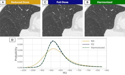
 CT images in a 56-year-old man with preexisting mild centrilobular emphysema at baseline who currently smoked. (A) CT scan imaged with a reduced-dose (RD) acquisition protocol and (B) full-dose (FD) acquisition protocol. (C) The harmonized image obtained with the noise-adaptive median filtering technique. (D) Corresponding lung density histogram.RSNA
Better quantifications of lung density -- enabled by harmonization techniques such as this one -- may produce more precise assessments of lung disease severity, according to the authors.
"Future research may be focused on the longitudinal utility of such a harmonization framework for providing local assessments of disease progression," they concluded.
Access the full study here.
Whether you are a professional looking for a new job or a representative of an organization who needs workforce solutions - we are here to help.