
The American College of Radiology (ACR) is rolling out the Pulmonary Embolism Reporting and Data Systems (PE-RADS) framework for diagnosing and classifying acute pulmonary embolism (PE) using CT and MR angiography.
Published July 21 in the RSNA journal Radiology and developed by the ACR, PE-RADS aims to streamline communication and patient care for acute PE.
"PE-RADS creates a common language and structured reporting system that can help radiologists and care teams communicate clearly, make informed treatment decisions, and align imaging findings with current clinical guidelines for the management of acute pulmonary embolism," said Lynne Koweek, MD, from Duke University in Durham, NC in a statement to AuntMinnie. Koweek chairs the ACR Committee on PE-RADS.
CT pulmonary angiography is the current primary method for diagnosing PE, though MR angiography is another suitable diagnostic test. However, images can be challenging for radiologists to interpret.
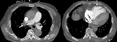
 Acute pulmonary embolism (PE) with right ventricle (RV) enlargement and clot-in-transit. PE chest axial CT angiography images demonstrate pulmonary embolus in the right pulmonary artery (white solid arrow), an enlarged RV (dotted arrow), and thrombus transiting through the right atrium (black
arrows), PE-RADS 4/RV+/T+. The “4” refers to the PE‑RADS ordinal category for the most proximal clot location (central arteries). “RV+” indicates RV enlargement (RV to left ventricle ratio ≥ 1.0). “T+” indicates definitive thrombus identified in the right atrium and/or RV.RSNA
The ACR developed PE-RADS version 2026 (v2026) with a zero-to-four grading scale and hierarchical framework to classify clot location combined with right heart imaging features:
0 – None
1 – Subsegmental
2 – Segmental
3 – Lobar or interlobar artery (past takeoff of upper lobe artery)
4 – Central (main, right, left)
N – Nondiagnostic study
PE-RADS aims to support risk stratification and guide patient management across multidisciplinary teams. PE-RADS also includes modifiers to indicate limitations in diagnosis stemming from image quality and non-PE causes of disease in the pulmonary arteries.
To develop the framework, the ACR and other societies nominated imaging specialists and clinicians from other specialties involved in PE care.
The ACR selected half of the specialists while the other half were nominated by the following medical societies: American College of Emergency Physicians, American College of Chest Physicians, Pulmonary Embolism Response Team Consortium, Society of Thoracic Radiology, Society for Cardiovascular Angiography and Interventions, and Society of Interventional Radiology.
The paper’s authors wrote that this framework aligns with existing PE guidelines and is designed for easy adoption and future expansion.
Koweek and colleagues highlighted that consistent terminology and reporting make way for effective communication between healthcare providers. It also "creates a stronger foundation" for studying outcomes, how resources are used, and future improvements in PE care.
"Our goal is to reduce reporting variability, improve patient care, and build a framework that supports better outcomes and more efficient use of health care resources,” Koweek told AuntMinnie.
The paper authors wrote that ongoing multicenter trials will help with refining management and more imaging findings that affect outcomes.
“The management of acute PE incorporates imaging findings, patient’s clinical status, and relevant risk factors, none of which alone can independently guide patient management,” they wrote.
Read the full paper here.
Whether you are a professional looking for a new job or a representative of an organization who needs workforce solutions - we are here to help.