


Virtual reality (VR) may have potential for supporting surgeons in evaluating abdominal neuroblastoma, suggest findings published October 15 in the Journal of Pediatric Surgery.
Compared with CT, VR improved surgeons’ anatomical assessment accuracy and confidence, wrote researchers led by Laura Privitera, PhD, from University College London Great Ormond Street Institute of Child Health in the UK.
“Overall, these findings support VR as a valuable complementary tool for anatomical assessment in abdominal neuroblastoma,” Privitera and colleagues wrote.
While CT improves visualization of tumor margins and their relationship to vital structures, challenges remain for preoperative evaluation of abdominal neuroblastomas. This is due to images representing complex 3D structures as stacks of 2D images, meaning surgeons must mentally reconstruct spatial relationships in 3D.
Emerging research suggests that VR improves preoperative planning and surgeon confidence and helps guide operative strategy in complex tumor resections. VR can help visualize tumor-vascular relationships and aid in decision-making.
The Privitera team studied whether patient-specific VR models improve the accuracy of preoperative anatomical assessment compared with conventional CT.
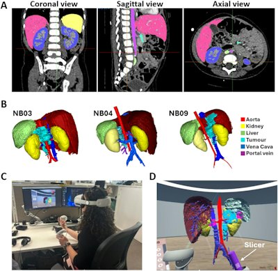
The team segmented CT scans from nine patients to create patient-specific VR models. From there, it imported the models into an in-house VR application. Eleven pediatric surgeons independently evaluated conventional CT images and VR reconstructions to assess vascular and organ involvement, image-defined risk factors, and operative strategies.
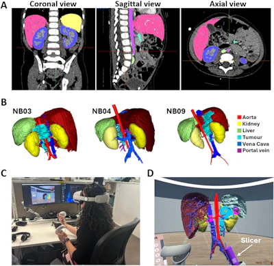 Workflow for generation and assessment of virtual reality (VR) neuroblastoma models. (A) Contrast-enhanced CT images of patient NB04 displayed in coronal, sagittal, and axial planes within the Materialise Mimics platform during the segmentation process. Segmented anatomical structures included the tumour, major vessels, kidneys, and liver. (B) Patient-specific 3D VR reconstructions generated for patients NB03, NB04, and NB09 following segmentation and post-processing. (C) A participant using the VR platform and headset to assess the patient-specific 3D reconstructions during the study session. (D) In-headset view of the VR environment demonstrating interactive manipulation of the 3D model using the slicer tool. Images are republished under a Creative Commons license (CC BY 4.0).Journal of Pediatric Surgery
VR led to an average increase of 12.4 percentage points for overall anatomical accuracy (p
VR did not lead to major changes in operative planning decisions or predicted tumor resectability. Surgeons said they had greater confidence in anatomical understanding and surgical planning with VR than CT (p
The researchers surveyed the surgeons to measure their attitudes toward the VR platform, using a 1-to-5-point Likert scale. High face validity ratings (scores 4 to 5) increased from 54/92 with CT to 82/86 with VR (p
The study authors highlighted the possibility of VR’s effects varying across specialties, considering radiologists typically interpret complex cross-sectional images.
“Further technological refinement, including improved segmentation and multimodal integration, together with larger prospective comparative studies are required to determine whether improved anatomical understanding translates into changes in operative decision-making or patient outcomes,” they wrote.
Read the full study here.
Whether you are a professional looking for a new job or a representative of an organization who needs workforce solutions - we are here to help.