
A team of researchers at Shenzhen Bay Laboratory in Shenzhen, China, has developed a wearable PET system capable of imaging the brain during free movement.
In a study involving phantoms and one human, the so-called “SmartBrain” system demonstrated spatial resolution and image quality comparable to a conventional clinical scanner, according to the group.
“The system’s lightweight, compact design and wearable configuration offer new opportunities for brain PET imaging beyond the constraints of conventional static systems,” noted lead author Han Liu, PhD, and colleagues. The study was published April 30 in the Journal of Nuclear Medicine.
Conventional brain PET requires patients to remain stationary in a sitting or supine position, posing particular challenges for children, patients with epilepsy, or others who cannot remain still, according to the authors. Conversely, wearable brain PET systems could extend metabolic imaging into natural and dynamic conditions, they suggested.
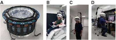
 (A) System comprises detectors and silicon photomultipliers with custom Hoffman brain phantom. (B) Seated-position scanning. (C) Backpack system permitting data collection in ambulatory states. (D) Workstation scenario demonstrating natural seated use.Journal of Nuclear MedicineTo that end, the group built SmartBrain. The system comprises a 16-sided polygonal ring with 192 detector modules arranged in six rings, using high-performance scintillator crystals coupled to silicon photomultipliers. The system weighs approximately 6 kg and can be worn via a backpack harness or suspension system. Two mechanical support configurations were developed to accommodate both seated and ambulatory use.
The group evaluated SmartBrain's physical performance against international standards (NEMA NU 2-2018) by conducting phantom studies using a custom Hoffman brain phantom and a multilayer Derenzo phantom. They also performed F-18 FDG imaging in a 43-year-old male patient with epilepsy, with results compared against a GE HealthCare Discovery MI PET/CT scanner.
In phantom testing, the system achieved a spatial resolution of 2.29 mm at the center of its field of view and resolved rod structures as small as 1.7 mm, which are performance metrics that meet requirements for human brain imaging, according to the investigators. In the human study, SmartBrain yielded cortical uptake patterns with well-defined gray matter distribution and preserved gyral anatomy that the authors described as comparable to the Discovery MI system.
“Although the sensitivity of the wearable system is lower than the DMI system, a 60-min scan acquired after low-dose F-18 FDG injection provided clear delineation of brain structures, with good comfort and feasibility during wear,” the group wrote.
The team wrote that the research remains at an early stage. Future optimization, including enhanced detector geometry, advanced correction algorithms, and AI-assisted image reconstruction, may further improve image quality, reduce acquisition times, and expand the scope of the system’s clinical and neuroscientific applications, it noted.
“The system enables ambulatory imaging with enhanced cost-efficiency and a space-saving design, positioning it as a scalable solution for precision diagnostics in both clinical and community health care settings,” the group concluded.
The full study is available here.
Whether you are a professional looking for a new job or a representative of an organization who needs workforce solutions - we are here to help.