
Radiologists, beware: CT imaging's diagnostic accuracy for identifying incidental adnexal lesions varies by finding type, according to a study published February 10 in Radiology.
The modality tends to correctly diagnose dermoids, simple cysts, and malignant ovarian lesions with metastases, but it is not as effective at identifying other types of these lesions -- suggesting that ultrasound and MRI may be necessary to correctly categorize CT adnexal findings, wrote a team led by Yang Guo, MD, of Brigham and Women's Hospital and Harvard Medical School in Boston.
"[Our study found that] all other adnexal lesions were challenging to diagnose at CT, and a substantial number of malignant lesions without metastases were mistaken for benign findings," the group noted.
How best to manage incidental adnexal lesions found on CT imaging depends on the diagnosis, but evidence is lacking regarding CT's performance for identifying these lesion types, the team noted. It wrote that "although CT is not considered the optimal modality for the characterization of an adnexal lesion, incidental lesions found at CT must be assessed to determine whether further characterization or follow-up is needed."
Guo's team evaluated interreader agreement and CT diagnosis of incidentally discovered adnexal lesions via a study that included data from 75 patients (mean age, 50 years) from between January 2022 and June 2023. Patients had been diagnosed at CT imaging with the following conditions:
Of these, 21% were malignant, according to the investigators.
Nine members of the Society of Abdominal Radiology Uterine and Ovarian Cancer Disease-Focused Panel -- blinded to the final diagnoses -- reviewed the CT images and used an American College of Radiology (ACR) white paper to determine the most likely diagnosis.
The researchers reported the following:
Reader performance for identifying adnexal lesions on CT imaging | ||
Type of lesion | Mean adjusted accuracy | Interreader agreement |
| Dermoids | 99% | 0.97 |
| Malignant ovarian lesions with metastases | 94% | 0.9 |
| Simple cysts | 86% | 0.64 |
| All other lesion types | 0.19 to 0.57 | |
They also found that overall, the nine readers more accurately diagnosed malignant lesions than benign lesions (82% vs. 52%, respectively; p
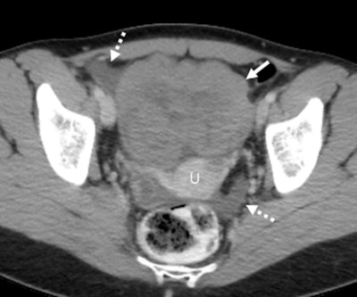
 Axial contrast-enhanced CT scan in a 28-year-old premenopausal woman shows an incidentally detected solid-appearing left ovarian lesion (solid arrow) anterior to the uterus (U), associated with trace ascites in the right adnexa and cul de sac (dashed arrows). At pathologic analysis, this was shown to be ovarian dysgerminoma. Two readers misdiagnosed this lesion, one as a leiomyoma and one as an ovarian fibroma. RSNA
The takeaway? "For other adnexal findings, ultrasound or MRI follow-up of the adnexal lesion found at CT is needed to support management recommendations," the group concluded.
Click here to access the full study.
Whether you are a professional looking for a new job or a representative of an organization who needs workforce solutions - we are here to help.